The images below represent many of the standard applications using Positron Emission Tomography (PET), Computed Tomography (CT), Fluorescence (FL), Bioluminescence (BLI), Near-infrared (NIR), Photoacoustic (PA), and Ultrasound (US) imaging. Not only do these images afford qualitative assessments, they also provide quantitative measurements to assess anatomical and functional processes over time. The SAIRF will train users on the software that is provided on our analysis workstations to generate graphics and quantify data such as those examples shown below.
Jump to:
CT Images
Bone Applications
Digitally Altered Femur
Monitoring Osteolytic Lesions Caused by Metastatic Breast Cancer
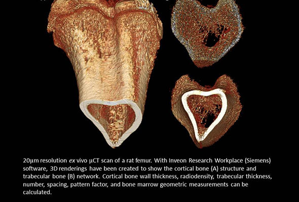
Trabecular Bone in Three Orientations
CT Scans and 3D Model of Mouse Heart/Aorta
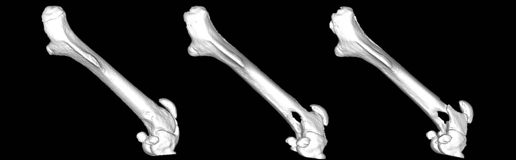
3D Femur Renderings
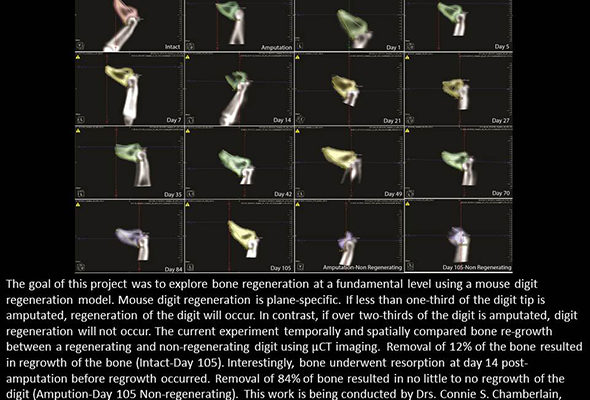
Bone Regeneration
Northern Cardinal - Density and Metabolic Modeling
CT Image of Metastatic Breast Cancer to Femur
Soft Tissue
Scan of a Mouse Injected with ExiTron Nano 12000 (Viscover ™)
Comparison of Scan with and without Contrast Agent Enhancement
Fenestra VC Enhanced
Frontal and Lateral View of Tumor
Fenestra LC to Assess Blood Supply
Finesta VC Scan
Fenestra VC Contrast versus No Contrast
3D Renderings and MIP Images from an in vivo μCT Scan
Transgenic PIRC Tumor Model
3D Rendering of Mouse Lungs
3D Rendering of a Mouse Lung
Intrapulmonary Shunt Vessels
3D Rendering of a Mouse Heart with μCT ex vivo.
in vivo μCT Scan of a Mouse with a Xenograft Tumor
Contrast Enhanced-CT - Prostate
Non-Traditional Applications
Wasp Identification
μCT used in Dendrochronology Applications
3D Printing a Prosthetic Hand
PET-CT images
PET Scan Versus Fused PET/CT Scan
microPET Scan of Glioblastoma Multiforme
Fused PET/CT in an Orthotopic Brian Tumor Model
Monoclonal antibody for imaging of brain malignancies
microPET Scan Targeting Nicotinic Receptor
microPET Scan - Prostate
Fused PET/CT Datasets
PET/CT image showing significant tracer uptake.
Uptake Comparison - Leukemia
Colon PET/CT Scan
Fused PET/CT Comparisons - Breast
64Cu-labeled TRC105-conjucated Hollw Mesoprous Silica Nanoparticles
Transgenic PIRC Tumor Model
Squamous Cell Carcinoma Xenograft
Optical Imaging
IVIS imaging of DsRed and GFP
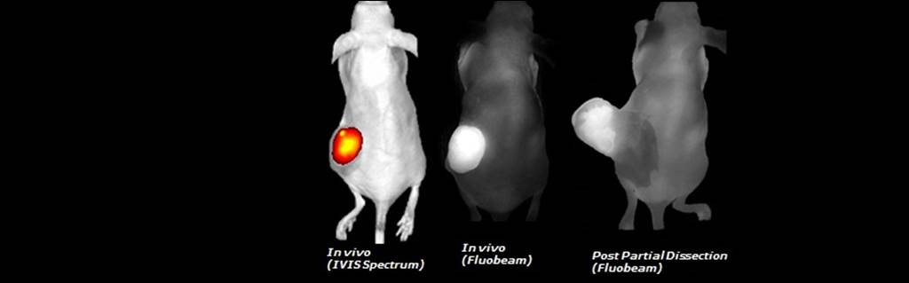
Comparison of IVIS and FluoBeam imaging modalities
Bioluminescent imaging of lung mets in a mouse model