The Imaging and Radiation Sciences (IR) Program pursues basic, translational, and clinical research involving ionizing and non-ionizing radiation in the diagnosis, staging, and treatment of cancer. The IR program has a strongly integrated scope in three major areas: Imaging Sciences, Radiation Sciences, and Imaging/Therapy Synergies.
Research Areas
The overarching goal of the IR program is to integrate physical and biological sciences with clinical research and clinical practice by developing and using novel imaging agents, imaging and therapeutic technologies, and radiobiologically and molecularly targeted approaches to improve cancer diagnosis and therapy.
- Aim 1: To develop and translate innovative imaging agents and technologies to improve cancer detection and treatment
- Aim 2: To develop molecular, immunological, radiobiological, and technological approaches that improve therapeutic radiation response
- Aim 3: To explore synergies between imaging and radiation sciences for more effective personalization of localized therapies
Imaging & Radiation Sciences Membership
View all Imaging and Radiation Sciences program members at UWCCC
Select Recent Accomplishments
This is an accordion element with a series of buttons that open and close related content panels.
Beau Biden Cancer Moonshot Initiative
A team of UW Carbone researchers led by Dr. Zachary Morris and Dr. Jamey Weichert was recently awarded $3.8 million from the NIH as part of the Beau Biden Cancer Moonshot Initiative. Funding from this award will support a multidisciplinary UW team that includes Drs. Bryan Bednarz, Paul Sondel, Mario Otto, David Vail, Mark Albertini, Randy Kimple, Kyungmann Kim, and Paul Harari. Together, these researchers and their respective labs will be evaluating whether a novel form of molecular targeted radiation therapy can modulate the tumor microenvironment in a way that promotes response to cancer immunotherapies.
Small Animal Imaging & Radiotherapy Facility (SAIRF)
The recent acquisition of two research radiation units represents a significant upgrade to the pre-clinical irradiation capabilities at UW. The small animal research radiation platform (SARRP) is capable of delivering intensity modulated radiation therapy (IMRT) using both coplanar and non-coplanar arcs in order to minimize normal tissue toxicity and better mimic clinical radiation delivery. These two units represent a significant investment by the School of Medicine and Public Health to support 21st century radiation delivery. Randy Kimple and Jason Timm led the efforts to upgrade these facilities at UW which was supported, in part, by a Department of Energy program focused on securing the radiation research infrastructure. This system is already being used by several NIH awards (PIs: Morris, Russell, Kimple) and multiple letters of support have been provided for future awards. Contact Justin Jeffery, SAIRF manager for more information.
Novel Molecular Imaging Agents
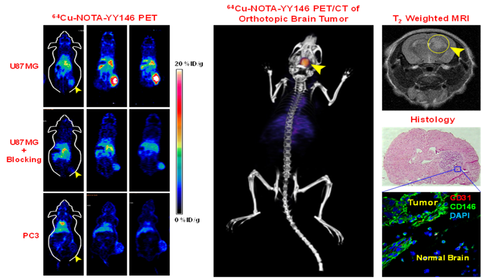
IR Program continues strong development of novel molecular imaging agents, particularly those related to imaging of angiogenesis (e.g., 89Zr-Df-TRC105) (Mol Pharm 20149) and novel agents for tumor detection of aggressive tumor phenotypes (Cai). For example, CD146 is a promising target for noninvasive in vivo imaging and targeted therapy of glioblastoma (e.g., 64Cu-NOTA-YY146) (Fig 1) Using YY146 as the primary antibody, histological studies of World Health Organization (WHO) grades I through IV primary gliomas showed positive correlation between CD146-positive staining and high tumor grade. In addition, multiple PET/MR and immuno-PET agents have been developed to allow combined whole body PET scanning to identify the location of tumor(s), followed up by the MR component (Bioconjug Chem 201410; Adv Mater 201411).
Novel Imaging Technologies
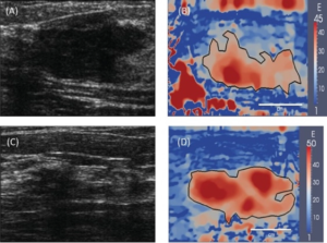
Young’s modulus images generated using elasticity imaging.
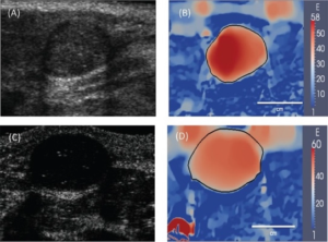
The Hall and Varghese groups have made outstanding progress in the development and refinement of quantitative methods in medical ultrasound. They have progressed the development of experimental methods for estimating specific parameters such as the acoustic backscatter coefficient (to quantify acoustic scattering on an absolute scale), effective scatter size (to describe the tissue microstructure), and the nonlinear elastic modulus (to describe the tissue stiffness on an absolute scale) (Ultrason Imaging 201412; J Pathol Inform 201413). These techniques have been translated into very productive intra-programmatic and inter-programmatic collaborations (TM members Keely, Eliceiri) studying breast (J Pathol Inform 201413; J Biomed Opt 201414) and cervical cancer structure (Ultrason Imaging 201215), particularly focusing on optical imaging techniques.
Imaging Biomarkers of Early Treatment Response
Jeraj continues the leading-edge research applying existing molecular imaging agents to novel and compelling oncologic applications, such as assessing [18F]FLT PET imaging (FLT is a marker of cellular proliferation) as a potential treatment response biomarker (Int J Radiat Oncol Biol Phys 20142.) Most notably, the UWCCC-funded pilot data on the use of FLT PET/CT in Acute Myeloid Leukemia (Leuk Res 201121,) has served as the key data for initiation of an ECOG-ACRIN EAI141 (NCT02392429) multi-center Phase II clinical trial: Early Assessment of Treatment Response in AML Using FLT PET/CT Imaging (Jeraj, Chair; Mattison (DT), Co-Chair) with the primary hypothesis that negative predictive value (NPV) of post-treatment FLT PET/CT assessment will be higher than NPV of post-treatment bone marrow biopsy. A key component of the EAI141 trial is incorporation of advanced Quantitative Total Marrow Imaging (QTMI) image processing tool, which allows for assessment of the whole bone marrow based on FLT PET/CT (Biomed Phys Eng Express 201622).