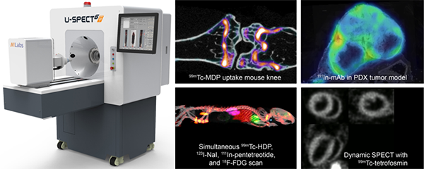
 The Small Animal Imaging & Radiotherapy Facility (SAIRF) was successfully awarded an NIH Shared Instrumentation Grant (SIG) S10 for an MILabs U-SPECT/CTUHR (ultra high-resolution) system. This system was installed in November, 2020 and is fully operational. This system, which is the only one of its kind in Wisconsin, allows for scanning animals up to the size of a large rat. Single Photon Emission Computed Tomography (SPECT) is a functional imaging technique that detects gamma rays resulting from radioactive decay from an injected radioisotope. A radioisotope can be chemically bound to a specific ligand that can then seek out and bind to a targeted biological marker. The U-SPECT from MILabs allows imaging of both traditional diagnostic imaging and therapeutic (theranostic) radioisotopes that are commonly used in brain, heart, bone and cancer applications, although many other uses exist as well. Computed Tomography (CT) is traditionally an anatomical imaging technique that uses x-rays to detect density. As such, CT is commonly used in bone applications, but it is also useful for lung and soft tissue applications when a contrast agent is administered. The MILabs CTUHR can achieve a resolution as good as ~15microns; a major upgrade to previous capabilities, which were limited to ~50microns. Please contact the SAIRF staff at SAIRF@uwcarbone.wisc.edu with any questions.