Small Animal Imaging and Radiotherapy Facility

University of Wisconsin–Madison Small Animal Imaging and Radiotherapy Facility (SAIRF) is a comprehensive core facility within the University of Wisconsin Carbone Cancer Center (UWCCC). We are dedicated to providing innovative, state-of-the-art, affordable, noninvasive, high-resolution, in-vivo and ex-vivo imaging and radiotherapy support to UWCCC investigators, University of Wisconsin researchers, and industry.
Small Animal Imaging and Radiotherapy Facility Logo Large

Leadership

Faculty Leader

Jamey P. Weichert

Facility Managers

Ashley Weichmann and Jenna Steiner
Viewing slide of 5
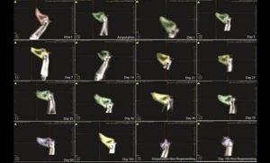
The goal of this project was to explore bone regeenration at a fundamental level using a mouse digit regeneration model. Mouse digit regeneration is plane-specific. If less than one-third of the digit tip is amputated, regeneration of the digit will occur. IN contrast, if over two-thirds of the digit is amputated, digit regeneration will not occure. The current experiment temporally and spatially compared bone re-growth between a regenerating and non-regenerating digit using μCT imaging. Removal of 12% of the bone resulted in regrowth of the bone (intact-Day 105). Interestingly, bone underwent resorption at day 14 post-amputation before regrowth occurred. Removal of 84% of bone resulted in little to no regrowth of the digit (Amputation-Day 105 Non-regenerating). This work is being conducted by Drs. Connie S. Chamberlain, Matthew A. Halanski, William L. Murphy, Xin Sun, Ray Vanderby, and students Lia Woods and Katie Swift.

Bone Applications

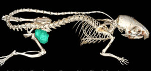
Routine in vivo μCT scan of a mouse with a xenograft tumor. This application can be used to make very accurate volume measurements such as tumor volume to assess treatment efficacy.

Soft Tissue

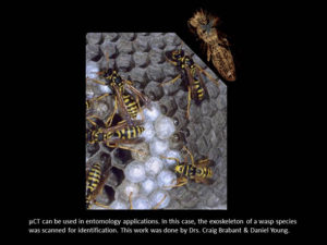
μCT can be used in entomology applications. In this case, the exoskeleton of a wasp species was scanned for identification. This work was done by Drs. Craig Brabant & Daniel Young.

Non-Traditional Applications

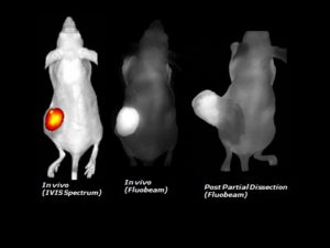
Comparison of IVIS and FluoBeam imaging modalities

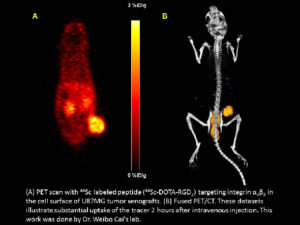
PET-CT images

(A) PeT scan with 44Sc labeled peptide targeting integrin αvβ3 in the cell surface of U87MG tumor xenografts. (B) Fused PET/CT. These datasets illustrate substantial update of the tracer 2 hours after intravenous injection. This work was done by Dr. Weibo Cai's lab.

Optical Imaging

Using the Facility

|

Any published research which received funding from the UWCCC Cancer Center Support Grant (CCSG), including the use of UWCCC Shared Resources to generate or analyze data or conduct clinical trials, must acknowledge the UWCCC. See Acknowledging UWCCC in Publications, Posters, and Presentations. Please acknowledge “UWCCC Circulating Biomarker Core” for any work supported by the CBC.  Thank you!

Sample Acknowledgment

“The author(s) thank the University of Wisconsin Carbone Cancer Center SAIRF, supported by P30 CA014520, for use of its facilities and services.”

Rates

The Small Animal Imaging and Radiotherapy Facility (SAIRF) is a fee-for-service laboratory available to UW Carbone Cancer Center (UWCCC) members, UW investigators, and industry. Contrast agents are not paid for by the SAIRF.

Contact SAIRF Staff to request a quote for PET, CT, Optical (fluorescence/bioluminescence), Hematologic, X-Ray, DXA, Ultrasound and Photoacoustic scanning. To request a quote for MRI, contact Beth Rauch.

Bill Pay

You can now pay by credit card using our Online Bill Pay option.

SAIRF Workflow

|

The facility director and manager of the Small Animal Imaging and Radiotherapy Facility (SAIRF) provide free consultation to determine which imaging and radiotherapy methodologies best meet the investigator’s research objectives.

  • Request consultation: Contact SAIRF Staff
  • Discuss logistics with SAIRF staff: animal protocols, housing, injection techniques, number of animals, time points, image resolution, tissue collection, contrast agents and modalities
  • For full-service imaging studies, scheduling can occur any time after initial consultation. Contact SAIRF Staff to coordinate full-service scheduling

  • Complete the Animal Transfer Form (pdf opens in new tab) one week prior to imaging. Transfer to SAIRF Umbrella Imaging Protocol (M005532; PI: Jamey Weichert; Rm: WIMR, B1334C). If needed, PIs can amend their protocol to include imaging procedures, SAIRF staff and room numbers
  • To comply with BioSafety communication standards, please fill out a Biohazard Intake Form as well
  • After approval, physically transport animals to B1334C housing
  • More information about animal housing in the SAIRF

  • General facility training (~30min session): Animal housing, PPE, radioactive and biohazard handling and disposal, etc. Additional equipment training is available upon request. Contact SAIRF Staff to schedule training
  • Send your full name and the 6-digit number on the back of your WisCard to the SAIRF Staff email to request key card access to the facility

  • For full-service, contact SAIRF Staff to coordinate scheduling
  • For self-service, after steps 1 through 3 are completed, schedule equipment directly in iLab.

  • Self-Service (users trained by SAIRF staff) or Full-service equipment:
    • Optical Imaging – fluorescence, bioluminescence, or near-infrared (IVIS, Fluobeam)
    • Animal and Cell Irradiators (Xstrahl CIX3, Xstrahl RS-225)
    • X-Ray & DXA Imaging (Faxitron)
    • Hematology Systems (HM5 and VS2)
  • Full-Service Equipment (operated by SAIRF staff only)
    • PET or PET/CT imaging (Inveon)
    • SPECT/CT (MILabs U-SPECT6CTUHR)
    • Ultrasound/Photoacoustic imaging (VevoLAZR 2100)
    • Targeted Animal Irradiator (Xstrahl SARRP)

  • Data is automatically backed up to UW’s ResearchDrive
  • Contact SAIRF Staff to schedule analysis training

  • The SAIRF can assist preparing images and videos, writing methods, as well as proposals/grant applications free of charge.

Select Publications

|

YearFirst AuthorArticleJournalDOI
2023Z. T. Rosenkrans, J. C. HsuAmplification of Cerenkov Luminescence Using Semiconducting Polymers for Cancer TheranosticsAdv Funct Mater10.1002/adfm.202302777
2023X. LiPretargeted Radioimmunotherapy of Ovarian Cancer with 225Ac and an Internalizing AntibodyJournal of Nucl Med10.2967/jnumed.123.266026
2023A. HoefgesAntibody landscape of C57BL/6 mice cured of B78 melanoma via a combined radiation and immunocytokine immunotherapy regimenFront Immunol10.3389/fimmu.2023.1221155
2023N. TsarovskyCyclophosphamide augments the efficacy of in situ vaccination in a mouse melanoma modelFront Oncol10.3389/fonc.2023.1200436
2023W. J. JinATM inhibition augments type I interferon response and antitumor T-cell immunity when combined with radiation therapy in murine tumor modelsJ Immunother Cancer10.1136/jitc-2023-007474
2023J. SunnerbergReview of optical reporters of radiation effects in vivo: tools to quantify improvements in radiation delivery techniqueJ Biomed Opt10.1117/1.JBO.28.8.080901
2023C. C. MitchellChanges in carotid artery texture by ultrasound and elastin features in a murine modelFront Cardiovasc Med10.3389/fcvm.2023.1215449
2023M. S. ReedMapping estimates of vascular permeability with a clinical indocyanine green fluorescence imaging system in experimental pancreatic adenocarcinoma tumorsJ Biomed Opt10.1117/1.JBO.28.7.076001
2023R. A. MukaddimIn Vivo Adaptive Bayesian Regularized Lagrangian Carotid Strain Imaging for Murine Carotid Arteries and Its Associations With Histological FindingsUltrasound Med Biol10.1016/j.ultrasmedbio.2023.05.017
2023S. L. V. HoffmanPreclinical studies of a PARP targeted, Meitner-Auger emitting, theranostic radiopharmaceutical for metastatic ovarian cancerNucl Med Biol10.1016/j.nucmedbio.2023.108368
2023K. A. O’LearyEstrogen receptor blockade and radiation therapy cooperate to enhance the response of immunologically cold ER+ breast cancer to immunotherapyBreast Cancer Res10.1186/s13058-023-01671-y
2023Y. ZhangIn Situ Vaccination Following Intratumoral Injection of IL2 and Poly-l-lysine/Iron Oxide/CpG Nanoparticles to a Radiated Tumor SiteACS Nano10.1021/acsnano.3c00418
2023M. M. ChoCD155 blockade enhances allogeneic natural killer cell-mediated antitumor response against osteosarcomabioRxiv10.1101/2023.06.07.544144
2023I. R. MarshTargeting of head and neck cancer by radioiodinated CLR1404 in murine xenograft tumor models with partial volume corrected theranostic dosimetryCancer Biother Radiopharm10.1089/cbr.2022.0084
2023W. LinTheranostic cobalt-55/58m for neurotensin receptor-mediated radiotherapy in vivo: A pilot study with dosimetryNuclear Medicine and Biology10.1016/j.nucmedbio.2023.108329
2023J. SoukupCorrelation of mineral density and elastic modulus of dog dentin using μ-CT and nanoindentationJournal of Biomechanics10.1016/j.jbiomech.2023.111434
2023J. SoukupMorphological quantification of the maxillary canine tooth in the domestic dog (Canis lupus familiaris)Annals of Anatomy10.1016/j.aanat.2022.152041
2023A. MuralidharTargeted Radiation and Immune Therapies—Advances and Opportunities for the Treatment of Prostate CancerPharmaceutics10.3390/pharmaceutics15010252
2023C. P. KerrDevelopments in Combining Targeted Radionuclide Therapies and Immunotherapies for Cancer TreatmentPharmaceutic10.3390/pharmaceutics15010128
2023P. M. CarlsonRadiation to all macroscopic sites of tumor permits greater systemic antitumor response to in situ vaccinationJ Immunother Cancer10.1136/jitc-2022-005463