TSB – Experimental Animal Pathology Laboratory
The Experimental Animal Pathology Laboratory (EAPL) provides a comprehensive array of histopathology services and expertise that enable UWCCC members to fully leverage the power of animal models of human cancer to reveal mechanisms that contribute to cancer development, progression, and responsiveness to therapies.
EAPL prioritizes 3 critical aims:
- Provide essential histology services to UWCCC members
- Facilitate the molecular analyses of normal and neoplastic tissues
- Provide histopathology expertise to UWCCC members and researchers
Experimental Animal Pathology performs histology services in their lab located on the fourth floor of the WIMR-I building.

Leadership
Supervisor
Dawn Church
Facility Staff
Joseph Hardin, Thomas Pier, Grant Mahant
Using the Facility
Provided by highly experienced senior histotechnologists, one junior histotechnologist and undergraduate students who have been trained by the senior histotechnologists.
Bring specimens to:
UWCCC Histology Lab
WIMR Room 4018
1111 Highland Ave.
Madison, WI 53705
- Preparation of histologic sections from both paraffin-embedded and frozen tissue, including serial sections
- Block preparations of cell pellets, embryoid bodies, and loose and agar embedded organoids
- Wide variety of routine and specialized histotechnology methods
- H&E
- Enzyme histochemistry
- Immunohistochemistry
- Special stains
- Slide preparation for in situ hybridization, LCM, and audioradiography
- Pathology consultation
Any published research which received funding from the UWCCC Cancer Center Support Grant (CCSG), including the use of UWCCC Shared Resources to generate or analyze data or conduct clinical trials, must reference the UWCCC. See Acknowledging UWCCC in Publications, Posters and Presentations. Please also acknowledge “UWCCC Experimental Animal Pathology Lab” for any work supported by the EAPL. Thank you!
Sample Acknowledgment
“The author(s) thank the University of Wisconsin Carbone Cancer Center Experimental Animal Pathology Laboratory supported by P30 CA014520, for use of its facilities and services.”
Rates are based on the service and client affiliation, as well as if the client needs the service performed on a ‘rush’ basis. UWCCC Members receive subsidized rates. Users outside of the UW should inquire for pricing.
Online Bill Pay
Pay by credit card using our Online Bill Pay option.
Scientific Accomplishments
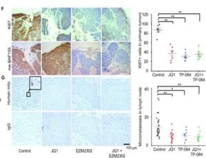
EAPL supported the work of Dr. Xu and colleagues who discovered methylated BAF155’s dual roles of hijacking super-enhancers and subverting anti-tumor activity to promote tumor metastasis.
EAPL staff processed, embedded, and generated serial sections of tumors, lymph nodes, and lung to support the analysis of proliferation and metastasis markers in experimental versus control mice in a patient derived xenograft model representing triple negative breast cancer.
Supported by R01 CA213293, R01 CA236356, IRA R35GM124806, Susan G. Komen PDF17481306
Published in Kim et al, Nucleic Acids Res. 2021 Dec 2;49(21):12211-12233. doi: 10.1093/nar/gkab1122. PMID: 34865122; PMCID: PMC8643633.
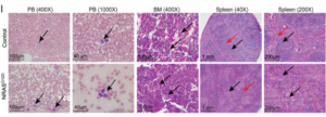
EAPL supported the work of Dr. Zhang and collaborators Drs. Patnaik, Nikolaev, et al, who demonstrated that RAS mutations drive proliferative chronic myelomonocytic leukemia (pCMML) via a KMT2A-PLK1 axis, which is a viable target for therapy against this aggressive subtype of CMML.
EAPL staff processed and embedded tissues, generated serial sections, and stained sections with H&E for histopathology analysis of the Vav-Cre-NrasG12D mouse model of pCMML compared to control mice.
Supported by R01CA152108, KL2 TR000136, CA136526
Published in Carr et al, Nat Commun. 2021 May 18;12(1):2901. doi: 10.1038/s41467-021-23186-w. PMID: 34006870; PMCID: PMC8131698.
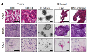
EAPL supported the work of Dr. Deming and colleagues who developed patient-derived cancer organoids across numerous cancer types that phenotypically represented the tissue of origin and successfully predicted, with bright-field and optical metabolic imaging technology, the response to treatment for a colorectal cancer patient.
EAPL staff processed and embedded patient-derived cancer organoids, generated serial sections, and stained sections with H&E for histopathology comparison to the tumor of origin.
Supported by R37 CA226526, R01CA205101, R01CA185747, R01CA211082, SU2C-AACR-IG-08–16, CBET-1642287
Published in Pasch et al, Clin Cancer Res. 25(17):5376-87, 2019. PMID: 31175091; PMCID: PMC6726566.
Location
Wisconsin Institutes for Medical Research (WIMR)
Lab: WIMR 4018
Office: WIMR 4003